Scandiflash x XRD:
Laboratory-Scale Flash X-Ray Diffraction
From seeing motion to understanding structural change, nanosecond Flash XRD without large-scale facilities.
Originaly published by Scandiflash, May 29, 2026
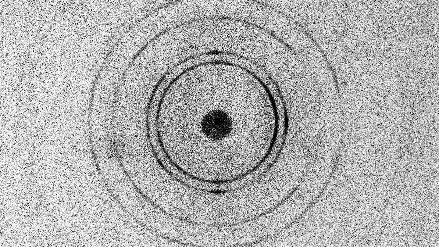
Scandiflash Flash XRD radiograph @ 25 ns.
Understanding Structural Change in Real Time
X-ray diffraction (XRD) is one of the most widely used methods for studying crystalline materials. Because diffraction probes atomic lattice structure directly, it provides quantitative information about phase composition, lattice strain, and crystallographic orientation (Figure 1).
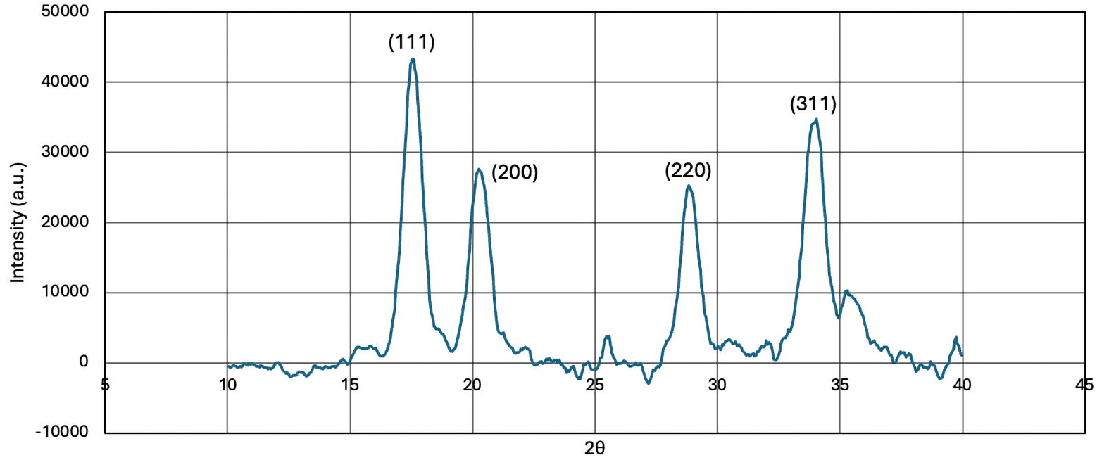
Figure 1. Raw XRD Curve
While conventional laboratory XRD systems operate under steady-state conditions, Flash X-ray diffraction enables measurements during transient events occurring on nanosecond to microsecond timescales. This makes Flash XRD a powerful diagnostic for dynamic materials research including shock compression, rapid heating, phase transitions, and high strain-rate deformation studies.
Flash XRD Advantages:
- Ultrafast pulse-resolved diffraction for dynamic processes
- Operando diffraction without pausing experiment
- Direct access to phase, strain & lattice evolution
- High-energy penetration in realistic sample geometries
- Precise timing & trigger-synchronized measurements
- Laboratory-scale alternative to synchrotron facilities
Example Applications:
- Phase transition kinetics
- Detection of combustion & chemical reaction
- X-ray diffraction as a thermodynamic diagnostic
- Dynamic compression studies
- Rapid heating experiments
- High strain-rate deformation analysis
Flash XRD Core Components:
- Flash X-ray source
- Electrodes
- X-ray optics & collimator
- Detector
- Shielding & safety enclosure
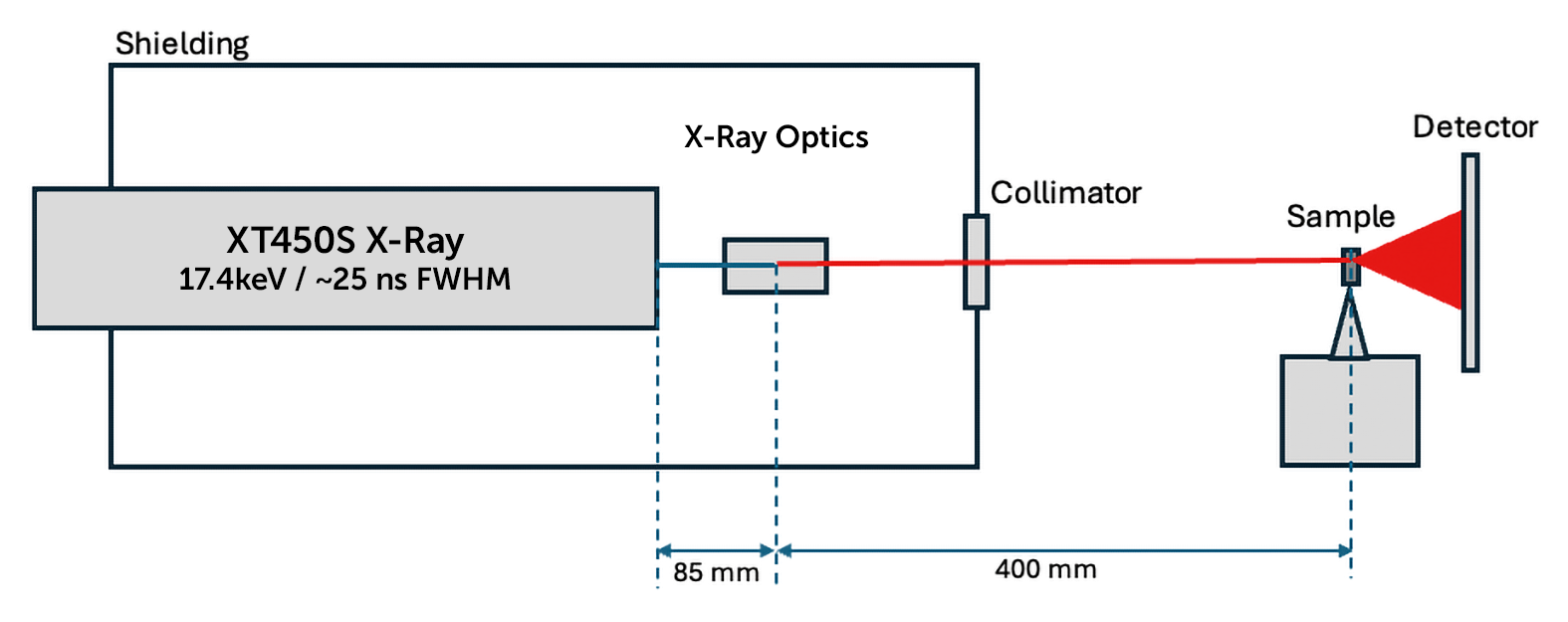
Figure 2. Flash XRD core components.
Structural Diagnostics During Dynamic Events
In a diffraction experiment, an incident X-ray beam interacts with lattice planes inside a crystalline material. When the Bragg condition is satisfied (Figure 3), diffraction peaks or rings are produced that correspond to specific crystallographic planes.
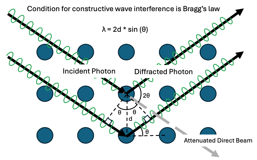
Figure 3. X-ray diffraction in crystal lattice.
Changes in diffraction peak position reveal changes in lattice spacing caused by compression, thermal expansion, elastic strain, and structural phase transitions. As a result, Flash XRD provides direct insight into the evolving thermodynamic and mechanical state of materials during dynamic experiments.
Time-Resolved Crystallographic Insight
Flash XRD combines short, intense X-ray pulses with diffraction geometry to resolve structural evolution during fast, non-equilibrium processes. Unlike conventional laboratory XRD, which often requires long integration times and interrupted experiments, Flash XRD enables measurements while the process remains fully active — preserving the true material response.
This allows researchers to:
- Capture structural evolution in real time
- Reveal transient & intermediate states
- Understand cause-and-effect relationships
- Study ultrafast material processes
- Validate theoretical & computational models
From Shock Physics to Operando Materials Research
Flash XRD is well suited for shock physics, detonation studies, impact research, and other extreme-condition applications where structural changes occur on nanosecond timescales.
The technique also extends naturally to slower time-critical systems such as batteries and electrochemical devices, where the most important structural changes often occur during short transient events within otherwise long experimental cycles. By enabling operando diffraction during continuous operation, Flash XRD captures processes that conventional laboratory techniques may miss entirely
Bridging the Gap Between Laboratory & Synchrotron XRD
Flash XRD bridges the gap between conventional laboratory diffraction and large-scale synchrotron experiments by combining crystallographic specificity with dramatically improved temporal resolution. The result is a compact laboratory-scale platform for dynamic diffraction studies that supports rapid experimental iteration, early-stage material screening, and synchronized multi-diagnostic research.
In many ways, Flash XRD is the structural analogue of high-speed imaging — not simply providing faster measurements, but enabling entirely new classes of observable material behavior.
Diffraction as a Thermodynamic Diagnostic
X-ray diffraction provides a non-contact diagnostic capable of inferring thermodynamic variables through measurements of lattice spacing. Compression shifts diffraction peaks toward higher 20 angles, while heating and expansion shift peaks toward lower angles (Figure 4). These peak shifts are governed by the material equation of state (EOS), enabling diffraction measurements to diagnose pressure, temperature, strain, and density evolution during dynamic experiments. ■
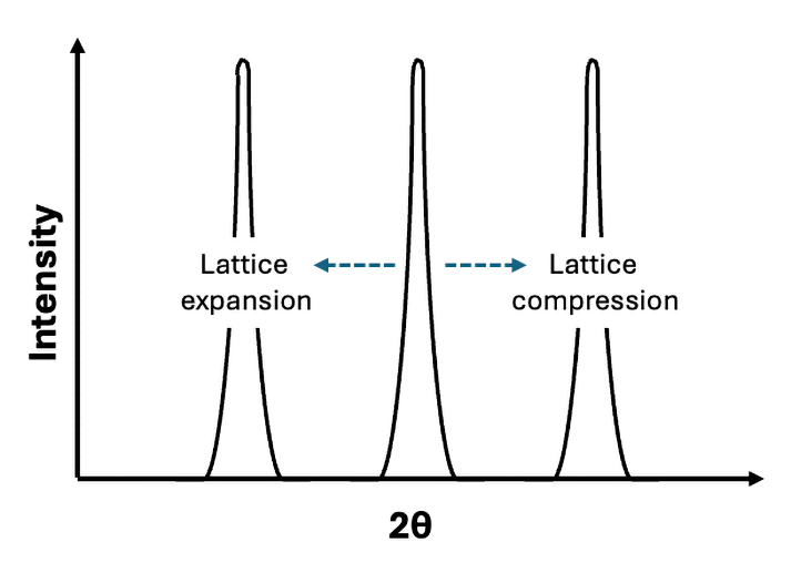
Figure 4. Lattice expansion/compression & diffraction peaks.
![]()
About Scandiflash
Scandiflash, located in Uppsala, Sweden, has been pioneering Flash X-ray technology for over 50 years – helping scientists and researchers around the globe see through the toughest conditions. Scandiflash Flash X-Ray (FXR) Systems are valuable solutions to image through fire, smoke, metal and other materials during research using fast 20 nanosecond X-ray pulses for incredibly sharp ultra high-speed imaging. Scandiflash FXR Systems are available for indoor lab setups and for outdoor large-scale firing ranges, and can be tailored to meet the requirements for a variety of research applications.
Learn more about ultra high-speed Flash X-ray imaging solutions:
Contact a Hadland Imaging representative to learn more about Flash X-ray imaging solutions & everything you need to get the job done right.
Keywords: Bragg’s Law, crystal lattice, Flash XRD, Laboratory-Scale Flash X-Ray Diffraction, Scandiflash, XRD