Shimadzu Application News
No: 01-01146-EN
March 2026
HyperVision HPV-X3 Ultra High-Speed Video Camera
Observation of Acrylic Block Fracture during Impact Compression Using the Hopkinson Bar Method
by Yuki Nishikawa
User Benefits:
- The Shimadzu HyperVision HPV -X3 offers three times the resolution of conventional models, enabling observation of specimen fracture at much higher resolution.
- With a maximum frame rate of 20 Mfps, the HPV-X3 is well-suited for observing high-speed phenomena such as an impact test.
Equipment:
- Shimadzu HyperVision HPV-X3 ultra high-speed video camera
- Nikon 105mm macro lens, bellows & close-up ring
- REL Split Hopkinson Pressure Bar/Kolsky Bar System
- Oxford Lasers FireFLY 500W Short-Pulsed Laser
Location: The Vault @ Hadland Imaging, R&D Training Center, Santa Cruz, California
Introduction
Understanding materials properties is critical to product design. In particular, for applications such as transportation equipment, where materials may be subjected to impact loads, it is essential to evaluate not only static properties but also impact properties. When subjected to impact, materials may exhibit stress and strain characteristics different from those under static loading, making it essential to quantify these characteristics through impact testing.
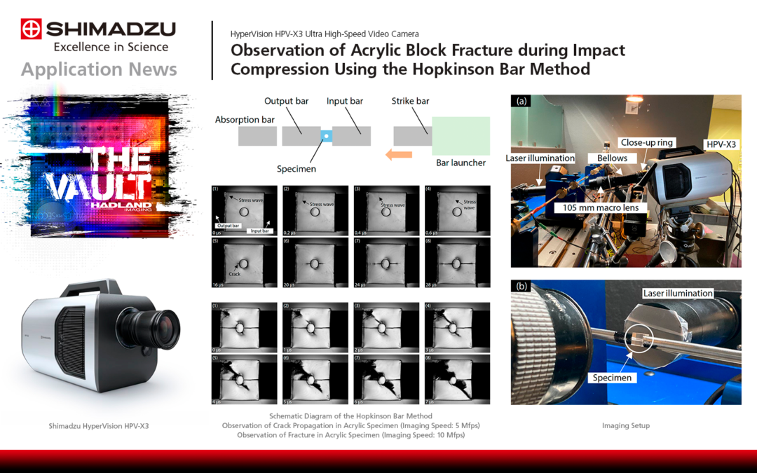
The Hopkinson bar method is a well-established technique for impact compression testing. Proposed by B. Hopkinson, this method enables detailed analysis of a material’s impact response and evaluation of its fracture properties by subjecting the specimen to a sudden force using a bar-launching device.
This paper presents an example of an impact compression test on an acrylic specimen with a central circular hole, recorded with an HPV-X3 high-speed video camera (Fig. 2) using the Hopkinson bar method.
Fig. 1 Schematic Diagram of the Hopkinson Bar Method
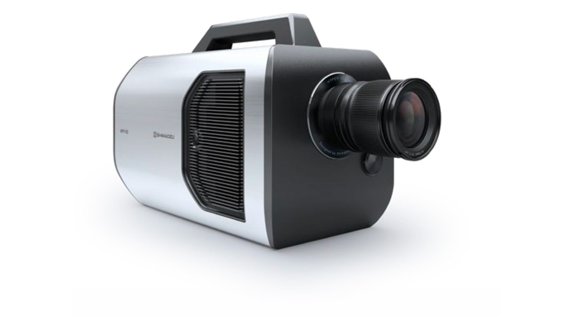
Fig. 2 Shimadzu HyperVision HPV-X3 Ultra High-Speed Video Camera
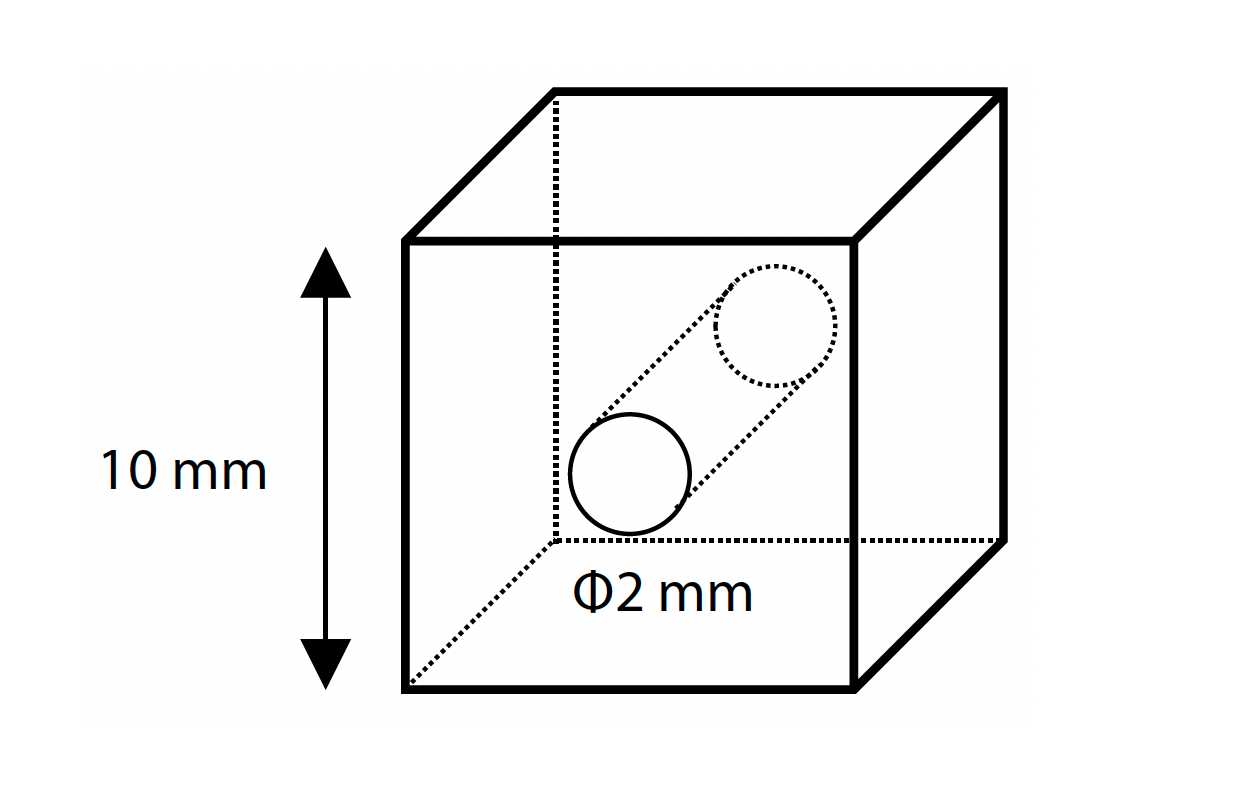
Fig. 3 Schematic Diagram of the Specimen
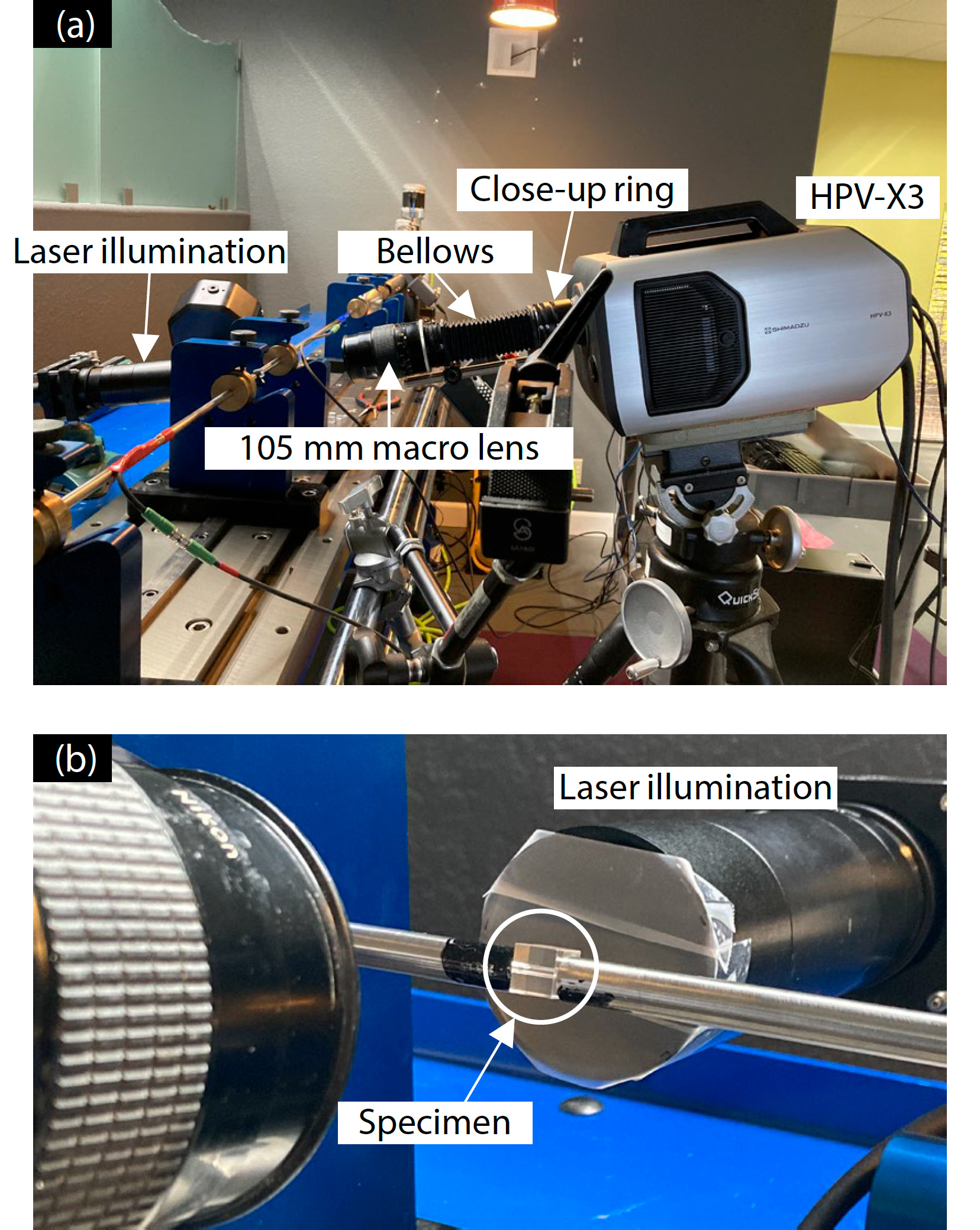
Fig. 4 Imaging Setup
| Table 1 Imaging Devices | |
| High-Speed Video Camera: | Shimadzu HyperVision HPV-X3 |
| Lens: | 105mm macro lens, bellows & close-up ring |
| Light: | Laser illumination |
| Table 2 Imaging Conditions | ||
| Recording Speed | Imaging Subject | |
| Imaging 1: | 5 Mfps | Immediately after loading Crack Propagation |
| Imaging 2: | 10 Mfps | Fracture Observation |
Imaging Results
The initial state of the acrylic specimen in Imaging 1 is shown in Fig. 5. Immediately after the specimen was subjected to impact compression by the input bar, a phenomenon believed to be a stress wave was observed. This phenomenon appeared in the upper left of the specimen in Fig. 5 (1), and subsequently, in (2) and (3), the wavefront was observed to propagate from left to right across the specimen. Later, in Fig. 5 (4), it can be seen that the wave, considered to be a stress wave, propagated to near the center of one side of the specimen. Based on this series of images, the speed of the stress wave was estimated to be approximately 2.7 km/s. Furthermore, in (5), which is 16 μs after Fig. 5 (1), a crack was initiated from the central circular hole of the specimen and propagated to the left, and from (6) to (8), it was observed to extend to both ends of the specimen’s side surfaces.
Next, the state of the specimen just before fracture in Imaging 2 is shown in Fig. 6. The crack propagation from the central circular hole to the left and right was similar to that observed in Imaging 1 (Fig. 5), but in Fig. 6 (1), the hole was compressed from both sides into an elliptical shape. Subsequently, crack propagation in the diagonal direction from the upper left of the specimen was observed. Similarly, crack propagation in the lower right of the specimen was also observed, and finally, complete fracture was confirmed. This fracture process was consistent with that reported in Related Application 3.
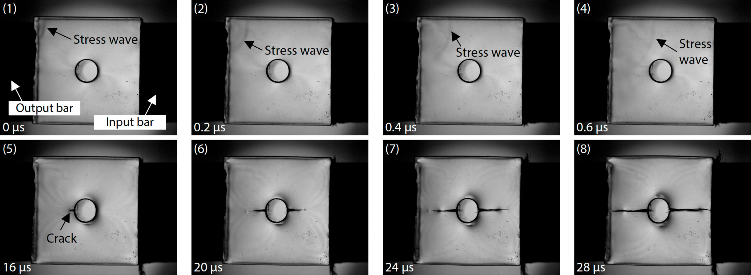
Fig. 5 Observation of Crack Propagation in Acrylic Specimen (Imaging Speed: 5 Mfps)
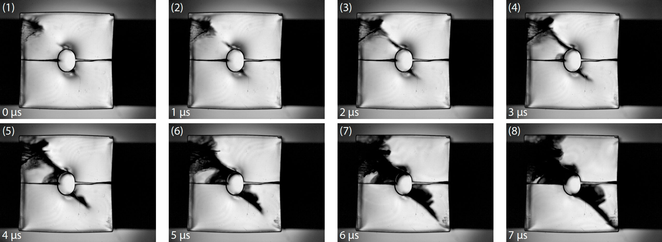
Fig. 6 Observation of Fracture in Acrylic Specimen (Imaging Speed: 10 Mfps)
Conclusion
The impact compression test of an acrylic specimen using the Hopkinson bar method was observed using the HPV-X3 ultra high-speed video camera. Inside the compressed specimen, the propagation of a wavefront believed to be a stress wave was first observed. Subsequently, cracks propagated from the central circular hole toward the left and right surfaces, and finally, diagonal cracks formed, leading to complete fracture. The progress of this series of fracture processes was clearly captured.
The HPV-X3 has three times the resolution of the previous HPV-X2 model, enabling more detailed observation of specimens during impact tests, including the Hopkinson bar method. Furthermore, because fracture phenomena during impact testing occur at high speeds, the HPV-X3, which can record images at up to 20 Mfps, is ideal for capturing them.
References
1) Hopkinson, B.: A Method of Measuring the Pressure produced in the Detonation of High Explosives or by the Impact of Bullets, Phil. Trans. Roy. Soc. Lond., A. 213, pp. 437-456, (1914).
Related Applications
- Observation of Fracture Behavior of Resin Material from an Impact Compression Test by the Hopkinson Bar Method, Application News No. V26
- 3D-DIC Analysis of a Metal Specimen Following an Impact Compression Test by the Hopkinson Bar Method, Application News No. V27
- Fracture Observation and Observation of Strain Distribution of Plastic Material with Hole in Impact Compression Test, Application News No. V29
![]()
Learn more about ultra high-speed visible, infrared, schlieren & Flash x-ray imaging solutions:
Contact a Hadland Imaging representative to learn more about the Shimadzu HyperVision HPV-X3 & The Vault @ Hadland Imaging, R&D Training Center in Santa Cruz, CA. Better Gear. Better Results. We’ve got the gear & knowledge you need to get the job done right!
Keywords: 20 million fps, impact compression testing, materials testing, pulsed laser illumination, REL Split Hopkinson Pressure Bar/Kolsky Bar System, Shimadzu Application News No: 01-01146-EN, Shimadzu HyperVision HPV-X2 ultra high-speed video camera, Shimadzu HyperVision HPV-X3 ultra high-speed video camera, The Vault at Hadland Imaging, Ultra High-Speed Imaging